
Breast cancer affects millions of women worldwide, and early detection remains one of the most important factors influencing treatment success and survival. In India, where cases are often diagnosed at younger ages and advanced stages, awareness of early warning signs and timely screening can make a significant difference.
That earlier onset is exactly why knowing the early signs and understanding what a mammogram can and can't tell you, matters more than ever. This guide explains breast cancer symptoms, mammogram screening, and the role of early detection in improving outcomes.
Why Early Detection Changes the Outcome
Breast cancer can often develop silently in its early stages, with many women experiencing no pain, visible changes, or noticeable symptoms. By the time warning signs become obvious, the cancer may have grown or spread, making treatment more complex. Early detection through regular screening and timely evaluation helps identify breast cancer when it is smaller and more manageable.
One of the most common mistakes is waiting until a lump, pain, or major breast change appears before seeking medical advice. However, many early breast cancers are detected through screening methods like mammograms before symptoms develop. Recognising changes early and following recommended screening schedules can improve treatment options and increase the chances of successful outcomes.
A breast change does not always mean cancer, but knowing which signs need medical attention can help with earlier diagnosis and better treatment decisions.
7 Early Signs of Breast Cancer You Shouldn't Ignore
Breast cancer symptoms can vary from person to person, and some changes may appear harmless at first. Many women delay evaluation because they associate symptoms with hormonal changes, infections, or normal body variations. However, recognising persistent or unusual changes and getting them checked early is an important step in breast health.
1. A new lump or thickening (and why most lumps are benign context, not alarm)
A new lump, hard area, or unusual thickening in the breast is one of the most recognised warning signs. However, it is important to understand that most breast lumps are not cancerous and can occur due to conditions like cysts, fibroadenomas, or hormonal changes.
The concern increases when a lump feels different from the surrounding tissue, remains over time, grows, feels hard, or is associated with other breast changes. Any new or unexplained lump should be evaluated by a healthcare professional rather than ignored.
2. Changes in breast size or shape
A sudden change in the size, contour, or shape of one breast may indicate an underlying change in breast tissue. While natural differences between breasts are common, a new asymmetry, unusual swelling, or change that develops without an obvious reason should be assessed.
Women often notice these changes while dressing, bathing, or during routine self-awareness checks, making awareness of what is normal for your body important.
3. Skin dimpling, puckering, or texture changes
Changes on the breast skin can sometimes indicate changes beneath the surface. Signs such as dimpling, puckering, thickened skin, or an “orange peel” texture (peau d’orange) should not be overlooked.
These changes may occur because cancer-related tissue growth can affect the skin’s attachment and appearance. Persistent skin changes, especially when they occur on one side, require medical evaluation.
4. Nipple changes; inversion, discharge, or scaling
New nipple changes can be an early warning sign. These may include:
- A nipple that turns inward when it was previously normal
- Unexplained discharge, especially bloody discharge
- Persistent crusting, scaling, or irritation around the nipple area
Not every nipple change indicates cancer, but new or unusual symptoms should be checked, particularly if they occur without injury or infection.
5. Persistent pain in one specific spot
Breast pain is commonly linked to hormonal changes, and pain alone is usually not the only sign of breast cancer. However, persistent pain that occurs in one specific area, does not improve, or feels different from usual breast discomfort should not be ignored.
A focused evaluation can help identify whether the pain is related to a benign condition or requires further investigation.
6. Swelling in the breast or underarm, even without a visible lump
Some breast cancers may cause swelling or fullness without creating an obvious lump. Changes may also appear in the underarm area due to enlarged lymph nodes.
Unexplained swelling, heaviness, tightness, or a new lump in the armpit area should be discussed with a healthcare provider, even if there is no visible breast abnormality.
7. Redness or unusual warmth (often mistaken for infection)
Redness, warmth, swelling, or tenderness of the breast can sometimes resemble an infection. While infections are a common cause, persistent symptoms that do not improve with treatment need further evaluation.
In clinical practice, breast specialists often see women delay consultation because symptoms like redness, swelling, or nipple changes are initially assumed to be due to infection or hormonal changes.
In Indian healthcare settings, another frequently missed sign is a painless lump, as many women wait for discomfort before seeking advice. Awareness and timely assessment remain important because early breast cancer may not cause significant pain or obvious symptoms.
Self-Exam vs Clinical Exam vs Mammogram: What's the Difference?
Detecting breast cancer early does not depend on a single method. Self-awareness, professional examination, and imaging tests each play a different role in identifying possible breast changes at different stages.
| Method | What It Can Detect | What It May Miss | Recommended Frequency |
|---|---|---|---|
| Breast Self-Exam / Breast Awareness | Helps women notice new lumps, changes in breast shape, nipple changes, or unusual symptoms | Cannot reliably detect very small cancers or changes deep inside breast tissue | Regular awareness of normal breast appearance and changes |
| Clinical Breast Exam (by a doctor) | A trained healthcare professional can identify suspicious lumps, skin changes, or other abnormalities | May not detect very small cancers that are not physically noticeable | Based on age, risk factors, and doctor’s recommendation |
| Mammogram Screening | Can detect early breast cancers, including small abnormalities and microcalcifications before symptoms appear | May require additional tests for unclear findings and is not suitable as the only evaluation method in every situation | Based on age, risk level, and screening guidelines |
Also Read: Recommended Cancer Screening Tests for Every Adults


Why none of the three should be used alone
Each screening approach provides different information. A self-exam helps women recognise changes early, but it cannot replace medical evaluation or imaging. A clinical breast exam allows specialists to assess abnormalities through physical examination, while a mammogram can detect changes that may not be felt by hand.
The most effective approach combines awareness of your own breast health, timely consultation with a healthcare professional, and recommended screening tests. Many early breast cancers may not cause noticeable symptoms, which is why relying only on how your breasts feel can delay diagnosis. Regular screening decisions should always be personalised based on age, family history, and individual risk factors.
A mammogram is one of the most important tools for detecting breast cancer early, but many women avoid it because they are unsure about the process. Understanding what happens during the test can make the experience easier and less stressful.
What Actually Happens During a Mammogram
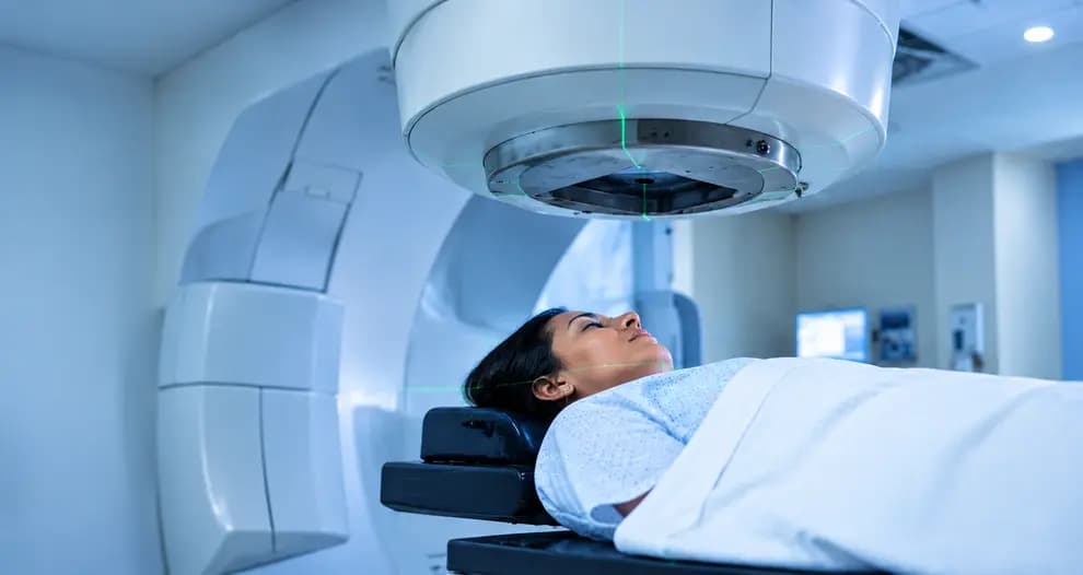
A mammogram is a specialised breast imaging test that uses low-dose X-rays to identify changes inside breast tissue, including abnormalities that may not be felt as a lump. The process is usually quick and involves a few simple steps.
Step-by-step: What to Expect During a Mammogram
1. Before the test
You will be asked about your medical history, previous breast problems, surgeries, or any symptoms you may have noticed. It is usually recommended to avoid applying deodorant, powder, or creams on the underarm and breast area on the day of the test, as these can sometimes affect image interpretation.
2. Breast positioning
During the procedure, a radiology technician positions each breast on the mammography machine. The breast is placed between two plates to capture clear images from different angles.
3. Compression of the breast
Compression gently flattens the breast tissue for a few seconds. This helps:
- Spread tissue evenly for clearer images
- Reduce movement-related blurring
- Lower the amount of radiation needed
Some women experience pressure or temporary discomfort during compression, but the process itself is very brief.
4. Duration of the test
A mammogram usually takes around 15–20 minutes, although the actual image capture time is much shorter.
2D vs 3D Mammogram (Tomosynthesis): What Is the Difference?
Traditional 2D mammography creates two-dimensional images of the breast. It is effective for routine screening but may be more challenging to interpret in women with dense breast tissue.
3D mammography (digital breast tomosynthesis) captures multiple images from different angles and creates a layered view of breast tissue. This can help radiologists examine areas more clearly, especially when overlapping tissue makes abnormalities harder to identify.
Breast density matters because dense breast tissue contains more glandular and connective tissue, which can sometimes make small cancers harder to detect on standard mammograms. Your doctor may recommend additional imaging depending on your breast density and risk profile
Understanding BI-RADS Score in Simple Terms
After a mammogram, the radiologist may use the BI-RADS (Breast Imaging Reporting and Data System) score to describe the findings and guide the next steps. A BI-RADS score does not automatically mean cancer. It is a standardised reporting system that helps doctors communicate results clearly. Common categories include:
- BI-RADS 0: More imaging is needed before a final assessment.
- BI-RADS 1: No abnormal findings; routine screening continues.
- BI-RADS 2: Benign (non-cancerous) findings.
- BI-RADS 3: Probably benign; follow-up imaging may be recommended.
- BI-RADS 4 or 5: Findings need further evaluation, often including a biopsy.
The purpose of BI-RADS is to ensure that women receive the right follow-up care based on their individual results.
Why Early Mammogram Access Matters
Breast cancer outcomes can differ significantly depending on when the disease is detected. However, access to screening and timely diagnosis is not equal everywhere. In many regions, including parts of India, women in rural areas may face challenges such as limited awareness, fewer screening facilities, travel barriers, and delayed specialist consultation compared with urban populations.
This difference can contribute to cancers being diagnosed at later stages in underserved areas, highlighting the importance of improving breast cancer awareness, screening access, and early medical evaluation.
A mammogram works best when combined with awareness of breast changes and regular medical guidance. It is not a replacement for clinical evaluation, but an important tool that can detect changes before they become noticeable.
Screening decisions are not the same for every woman, and the right time to begin depends on age, personal risk factors, and regional cancer patterns. Understanding the difference between global recommendations and India-specific considerations can help women make informed decisions.
When Should You Start Mammogram Screening? (India vs Global Guidelines)
There is no single screening age that applies to every woman. Guidelines are developed based on population risk, healthcare access, and patterns of when breast cancer commonly occurs. While many international recommendations focus on screening from the 40s onward, breast cancer patterns in India have led many specialists to emphasise earlier awareness and personalised screening decisions.
US Guidelines vs India-Specific Considerations
In countries like the United States, organisations such as the American Cancer Society and United States Preventive Services Task Force generally recommend that women begin regular mammogram screening around age 40, with the frequency depending on age, risk, and individual preference.
In India, many breast specialists consider the fact that breast cancer is often diagnosed at a younger age compared with Western populations. While routine screening recommendations may vary, oncologists commonly focus on:
- Starting conversations about breast health earlier
- Assessing individual risk factors instead of following age alone
- Encouraging timely evaluation of any suspicious breast changes
This is especially important because some women in India may delay consultation until symptoms become noticeable, which can affect the stage at diagnosis.
Who May Need Earlier Mammogram Screening?
Women with higher-than-average risk may require earlier or additional screening methods based on specialist advice.
1. Family History of Breast Cancer
Women with a mother, sister, or close relative diagnosed with breast cancer may have an increased risk. Doctors may recommend earlier screening, genetic counselling, or additional imaging depending on the family history pattern.
2. BRCA1 and BRCA2 Gene Mutations
Inherited changes in genes such as BRCA1 and BRCA2 can significantly increase breast cancer risk. Women with these mutations often follow a more intensive screening plan, which may include starting earlier than standard recommendations and using additional imaging methods.
3. Dense Breast Tissue
Dense breasts contain more glandular tissue compared with fatty tissue. This can:
- Make some cancers harder to identify on mammograms
- Increase breast cancer risk in some women A doctor may recommend additional evaluation, such as ultrasound or MRI, depending on overall risk.
4. Previous Chest Radiation Exposure
Women who received radiation therapy to the chest area at a younger age, particularly for conditions such as certain cancers, may have a higher future breast cancer risk. These individuals may need a personalised screening approach.
**Also Read: Which Is the Best Oncologist in Kozhikode for Expert Cancer Treatment? ** Misinformation around mammograms often prevents women from getting timely screening. Understanding the facts can help women make better decisions about breast health.
Common Myths About Mammograms, Addressed Plainly
Many concerns about mammograms come from misinformation or personal experiences shared without medical context. Let’s look at some of the most common myths and what the evidence actually shows.
“Mammograms cause cancer”
A common concern is that radiation from mammograms may cause cancer. However, mammograms use a very low dose of radiation, and the benefits of detecting breast cancer early generally outweigh the minimal radiation exposure involved. The test is carefully regulated and performed using safety standards.
“If it doesn't run in my family, I'm safe”
Family history is only one risk factor. Many women diagnosed with breast cancer do not have a known family history. Age, hormonal factors, lifestyle, and other biological factors can also influence risk.
“Mammograms hurt too much to be worth it”
Some women experience temporary pressure or discomfort during breast compression, but the procedure is usually completed within minutes. The brief discomfort helps create clearer images and improves detection accuracy.
“Young women don't need screening”
While routine screening schedules often begin at specific ages, younger women can still develop breast cancer. Those with symptoms, strong family history, genetic risk, or other risk factors may need earlier evaluation.
Early breast cancer detection is not only about having access to screening technology but also about accurate interpretation, timely diagnosis, and coordinated specialist care. A comprehensive approach helps patients move from detection to treatment planning without unnecessary delays.
How Baby Memorial Hospital Approaches Early Breast Cancer Detection
Baby Memorial Hospital focuses on early breast cancer detection through a combination of advanced imaging, specialist expertise, and multidisciplinary care.
-
Advanced breast imaging: The hospital uses digital mammography with 3D tomosynthesis, which provides detailed layered images of breast tissue. This can help improve detection accuracy, especially in women with dense breast tissue where abnormalities may be harder to identify.
-
Multidisciplinary cancer care: Breast health evaluation involves collaboration between radiologists, surgical oncologists, and cancer care specialists, supported through the American Oncology Institute partnership. This ensures findings can be reviewed and managed through a coordinated care pathway.
-
Better diagnostic decision-making: Combining advanced imaging with specialist review can help reduce unnecessary recalls, improve evaluation of suspicious findings, and support personalised treatment planning when required.
-
Accessible expert care: Serving patients from across Kerala, other parts of India, and international locations, Baby Memorial Hospital provides access to advanced breast cancer screening and specialist consultation while maintaining structured diagnostic standards.
For women with symptoms, family history, or concerns about breast cancer risk, timely screening and consultation with the right specialists can help identify problems earlier and support better treatment outcomes.
Take your NAVAyug Cancer screening test today at BMH.
A breast change or an abnormal mammogram result can feel concerning, but it does not always mean cancer. The next steps focus on getting clear answers through proper evaluation rather than assuming the worst.
What to Do If You Notice a Symptom or Get an Abnormal Mammogram Result
If you notice a new lump, breast change, or receive an abnormal mammogram report, avoid delaying follow-up. Many findings turn out to be benign, but timely assessment helps identify the cause.
- Consult a breast specialist: Discuss your symptoms or mammogram findings with a qualified doctor.
- Additional imaging if needed: A diagnostic mammogram, breast ultrasound, or other imaging tests may be recommended for a closer evaluation.
- Biopsy when required: If imaging shows a suspicious area, a small tissue sample (biopsy) may be performed to confirm the diagnosis.
Early evaluation provides clarity and helps ensure the right treatment approach if further care is needed.
Connect with a medical assistant at Baby Memorial Hospital for guidance on screening, consultations, and the right care pathway.
Conclusion
Breast cancer may develop silently, but recognising early signs and following recommended screening practices can help identify changes at a more manageable stage. Mammograms, clinical evaluations, and awareness of breast changes each play an important role in early detection. If you notice any unusual symptoms or receive an abnormal screening result, timely consultation can provide clarity and guide the next steps.
At Baby Memorial Hospital, patients can access advanced breast imaging, specialist evaluation, and coordinated cancer care support. Chat with BMH’s medical assistant today to understand your screening and consultation options.
Medical Disclaimer: This article is intended for general informational purposes only and does not constitute medical advice, diagnosis, or treatment. Always consult a qualified physician or oncologist regarding any symptoms, screening schedule, or treatment decisions specific to your health. If you notice any of the signs discussed above, seek prompt evaluation from a licensed healthcare provider.