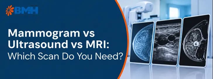

Choosing the right breast screening method can feel overwhelming when you're trying to protect your health. Mammograms, ultrasounds, and MRI scans each offer different advantages depending on your age, risk level, and breast tissue type. They are not competing tests. Each one sees breast tissue differently, so the right choice depends on your age, breast density, family history and whether you have a symptom. This guide explains what each scan does, who needs which one, and what actually happens on the day, drawing on the breast cancer care pathway at Baby Memorial Hospital (BMH), Kozhikode.
Quick answer: A mammogram is the standard screening test for average-risk women from age 40. An ultrasound is a radiation-free add-on for dense breasts, lumps, young women and pregnancy. An MRI is the most sensitive scan, used for high-risk women (BRCA mutation or roughly 20-25% lifetime risk) and for unclear findings. Doctors often combine them.
How Each Breast Scan Works
Mammograms, ultrasounds and MRIs are three distinct breast imaging tools. Here is each one in plain English.
What Is a Mammogram?
-
How it works: low-dose X-rays image the breast. Options include 2D mammography and 3D tomosynthesis, which improves detection and reduces callbacks.
-
Primary use: the gold-standard screening test for average-risk women from 40. The American College of Radiology and American Society of Breast Surgeons advise yearly screening from 40; the American Cancer Society suggests yearly at 45-54, then yearly or every two years, as summarised in this guideline review. The National Comprehensive Cancer Network also advises yearly mammograms from 40.
-
Best at detecting: tiny calcium deposits (microcalcifications), distortions in tissue structure and overall changes across the whole breast.
-
Limitations: minor ionising radiation, and dense breast tissue can mask tumours or lower sensitivity.
What Is a Breast Ultrasound?
-
How it works: high-frequency sound waves, with no radiation, create real-time images inside the breast.
-
Primary use: a supplemental screening or diagnostic tool, often used to follow up a suspicious lump on a mammogram or to assess dense breasts. It is also the first-line choice for patients under 30 and pregnant women because there is no radiation.
-
Best at detecting: whether a lump is a solid tumour or a fluid-filled cyst. Automated breast ultrasound (ABUS) is one option for dense tissue.
-
Limitations: it cannot see microcalcifications well, depends on the operator’s skill and has a higher false-positive rate.
What Is a Breast MRI?
-
How it works: powerful magnets, radio waves and an intravenous contrast dye (gadolinium).
-
Primary use: high-sensitivity screening for high-risk women, such as BRCA carriers or those with a 20-25% or higher lifetime risk, typically from about age 30, and for clarifying unclear results. The American Cancer Society’s expert panel recommends yearly MRI alongside mammography at that risk level. MRI is added to, not a replacement for, mammography.
-
Best at detecting: soft-tissue detail and abnormal blood-vessel patterns. Breast density does not affect it.
-
Limitations: much more expensive, longer, less able to show microcalcifications and prone to false positives.
Mammogram vs Ultrasound vs MRI: Side-by-Side Comparison
| Feature | Mammogram | Ultrasound | MRI |
|---|---|---|---|
| Technology | Low-dose X-rays | Sound waves | Magnets, radio waves and contrast dye |
| Radiation | Yes (low dose) | No | No |
| Primary role | Standard yearly screening (age 40+) | Diagnostic follow-up; dense-breast add-on | High-risk screening; problem-solving |
| Best for | Calcifications and overall architecture | Cysts versus solid masses | High-risk women and dense-tissue detail |
| Relative cost | Moderate | Lower than a mammogram | Highest |
| Main weakness | Dense tissue can hide tumours | Misses microcalcifications; operator-dependent | Cost, time and false positives |
Cost ranking is general: ultrasound is cheaper than a mammogram, while MRI’s higher cost and false-positive rate are why researchers hesitate to recommend it for women at only moderately raised risk.
What Actually Happens on the Day
Patients often tell us the waiting and the unknown worry them more than the scan. These practical points come from the imaging routines doctors commonly follow.
Mammogram
- Wear a two-piece outfit. Skip deodorant, powder and lotion on the chest and underarms, as they can show up as bright specks on the image.
- Carry old reports and films. Radiologists compare year to year, and a stable finding is reassuring.
- Each breast is pressed between two plates for a few seconds per view. The pressure is firm and uncomfortable, but brief. Booking the week after your period usually means less tenderness.
- Tell the technologist about implants, a possible pregnancy and any lump you have noticed.
Ultrasound
- No preparation. Gel is applied and a hand-held probe is moved over the breast and underarm.
- Point to the exact spot you are worried about. Because the result depends on the operator, a lump you can feel should always be shown to the person scanning.
MRI
- You lie face down on a padded table with openings for the breasts. A contrast dye is given through a vein, and the machine makes loud tapping sounds, so earplugs or headphones help.
- Tell the team about a pacemaker, metal implants, kidney problems, pregnancy and any fear of enclosed spaces before the day.
- Premenopausal women are often scheduled in the second week of the menstrual cycle, so ask the imaging team which day suits you.


Why Do Patients Choose BMH for Breast Cancer Care?
Breast cancer care involves more than a scan or a single consultation. Patients may need diagnostic imaging, a biopsy, pathology review, surgery, drug therapy or radiation, depending on their diagnosis. Having these services coordinated through one hospital can make the process easier to navigate.
At Baby Memorial Hospital (BMH), Kozhikode, the NAVA Cancer Institute brings together multiple oncology specialties and diagnostic services to support patients through different stages of cancer care.
What Makes a Difference in Breast Cancer Care?
1. Diagnostic services that support timely decisions
An abnormal mammogram or breast scan does not automatically mean cancer. Further assessment may involve additional imaging and a biopsy, followed by a pathology report. BMH has PET-CT, radiology and pathology facilities to support the diagnostic process.
2. Access to different treatment options
Breast cancer treatment depends on the type, stage and characteristics of the tumour, as well as the patient's overall health. BMH's breast cancer services include surgical oncology and TrueBeam LINAC radiotherapy, alongside medical and radiation oncology expertise.
Depending on the individual case, treatment planning may involve:
-
Breast-conserving surgery or oncoplastic surgery
-
Chemotherapy, hormonal therapy or other appropriate systemic treatments
-
External beam radiation therapy
-
Selected specialised radiation approaches, where clinically suitable
3. A coordinated approach to treatment
Breast cancer decisions often involve more than one specialist. Coordination between medical, surgical and radiation oncologists, radiologists and pathologists helps the team interpret findings and plan the next steps. BMH's medical oncology services describe multidisciplinary discussions and the use of diagnostic and molecular information to guide treatment decisions.
4. Support for patients travelling for care
Patients coming to Kozhikode from other parts of India or abroad may need help coordinating appointments, medical records and treatment estimates. BMH states that it supports national and international patients through dedicated case management and multilingual assistance.
Before travelling, patients should confirm appointment availability, the documents required for review and the expected costs. A written estimate can help families plan for consultations, investigations and treatment.
BMH's NAVA Cancer Institute is one option patients can consider when seeking coordinated breast cancer evaluation and treatment in Kozhikode. The most appropriate centre will depend on the patient's diagnosis, clinical needs, location and budget.
If you already have a mammogram, ultrasound, biopsy report or other investigation results, arrange a specialist consultation to understand what they mean and whether further assessment is needed. An abnormal scan alone is not a confirmed cancer diagnosis, and the next step should be guided by a qualified clinician.
Conclusion
Mammogram, ultrasound and MRI are teammates. Start with a yearly mammogram from 40 if your risk is average, add an ultrasound if your breasts are dense or you have a lump, and talk to your doctor about MRI if a BRCA mutation or strong family history puts your lifetime risk near 20% or higher. Younger Indian women, who are diagnosed earlier and rarely screened, should not ignore a lump just because they are under 40. The scan is only the first step.
If your report raises questions, book a consultation with the breast care team at BMH, Kozhikode, to review your images and plan what comes next.
Medical disclaimer: This article is for general information and does not replace advice from a qualified doctor. Screening ages, scan choices and timing depend on your personal and family history. Please consult a doctor before making decisions about tests or treatment.