
Vulvar cancer is a rare cancer affecting the external female genitalia, and its early symptoms can resemble common problems such as irritation, itching, or skin changes. Knowing which symptoms need medical evaluation can help detect the disease before it spreads. This guide explains the key vulvar cancer symptoms, including persistent itching, pain, bleeding, lumps, sores, and changes in skin colour or texture.
It also covers the possible causes and risk factors, how doctors diagnose and stage vulvar cancer, and the treatment options available at different stages. You’ll also learn about survival rates and what current data shows about outcomes when vulvar cancer is diagnosed early.
What Is Vulvar Cancer?
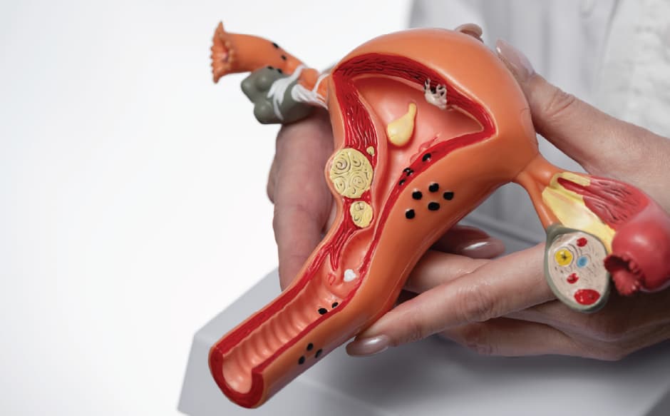
The vulva is the external part of the female genitalia, it includes the labia majora (outer lips), labia minora (inner lips), the clitoris, and the vaginal opening. Vulvar cancer develops when cells in this skin and tissue begin growing abnormally and uncontrollably.
It's frequently confused with two other gynaecologic cancers:
| Cancer type | Where it starts | Key distinguishing point |
|---|---|---|
| Vulvar cancer | Outer genital skin (labia, clitoris, perineum) | Usually visible/palpable on the skin surface |
| Vaginal cancer | Inner vaginal canal | Rarely visible without internal exam |
| Cervical cancer | Cervix (opening of the uterus) | Detected via Pap smear/HPV test, not visible externally |
Early Signs and Symptoms of Vulvar Cancer
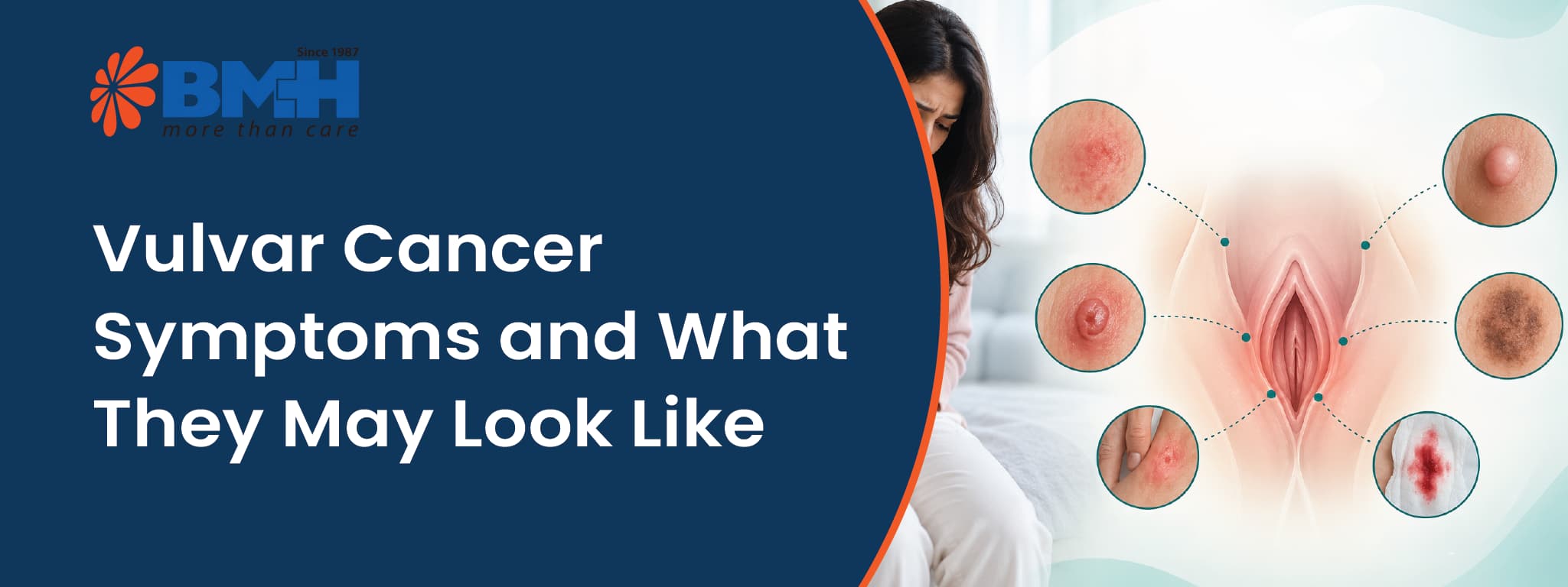
Because early vulvar cancer often looks like a minor skin issue, it's commonly self-treated with over-the-counter creams for weeks or months before a doctor sees it. The symptoms to actually take seriously if they persist beyond 3-4 weeks:
| Symptom | What it may look/feel like |
|---|---|
| Persistent itching | Doesn't resolve with usual creams or hygiene changes |
| Skin colour or texture change | A patch that's lighter, darker, thicker, or wart-like |
| A lump or open sore | May bleed, ooze, or not heal like a normal cut |
| Pain or burning | Especially during urination or intercourse |
| Unusual bleeding or discharge | Not related to a menstrual cycle |
| A mole that changes | New asymmetry, colour change, or growth on the vulva |
Many of these symptoms overlap with non-cancerous conditions like lichen sclerosus, yeast infections, or dermatitis, which is exactly why a biopsy, not a guess, is the only way to tell the difference.
What Causes Vulvar Cancer? Key Risk Factors
Vulvar cancer doesn't have one single cause, it generally develops along one of two distinct biological pathways.
HPV infection.
Certain high-risk strains of human papillomavirus, especially HPV-16, are linked to a large share of vulvar cancers, particularly in younger women. This is the same virus responsible for most cervical cancers, which is why HPV vaccination is considered a genuine prevention tool, not just a cervical-cancer measure.
Lichen sclerosus and VIN.
In older women, vulvar cancer more often develops from long-standing lichen sclerosus (a chronic skin condition causing thinning, itching, and white patches) or vulvar intraepithelial neoplasia (VIN), a pre-cancerous change in the surface cells. About 4% of women with lichen sclerosus eventually develop vulvar cancer, which is why this condition is not something to simply manage with steroid creams and ignore long-term.
Other risk factors include increasing age (most diagnoses occur after 50), smoking, a weakened immune system (including HIV or long-term immunosuppressant use), and a history of cervical or vaginal pre-cancer.
How Vulvar Cancer Is Diagnosed
A visual exam alone can't confirm vulvar cancer, the process usually looks like this:
-
Clinical examination: A gynaecologist inspects the vulva for suspicious lesions, colour changes, or growths.
-
Colposcopy: A magnifying instrument examines the vulvar skin in detail if the abnormality isn't clearly defined.
-
Biopsy: A small tissue sample is removed and examined under a microscope; this is the only definitive way to confirm cancer and its subtype.
-
Imaging (CT/MRI/PET): Ordered if there's concern the cancer may have spread to lymph nodes or beyond, to help plan treatment.
Also Read: PET Scan vs CT Scan vs MRI for Cancer: Which Is Right for You?


Stages of Vulvar Cancer and Survival by Stage
Staging determines both treatment intensity and prognosis. Vulvar cancer is staged using the FIGO/AJCC system, but for prognosis purposes, it's often simplified into three groups:
| Stage | Description | 5-Year Relative Survival |
|---|---|---|
| Localized | Confined to the vulva; no spread to lymph nodes | 86% |
| Regional | Spread to nearby tissue or lymph nodes (groin) | 53% |
| Distant | Spread to distant organs (lungs, liver, bone) | 19% |
Vulvar Cancer Treatment Options
Vulvar cancer treatment depends mainly on how far the cancer has spread, along with the tumour’s size, location, and lymph node involvement.
-
Wide local excision: Removal of the tumour with a margin of healthy tissue, used for small, early-stage lesions where preserving vulvar anatomy is possible.
-
Vulvectomy: Partial or complete removal of the vulva for larger or more advanced tumours.
-
Groin lymph node evaluation: Sentinel lymph node biopsy or full groin lymph node dissection to check for spread, since node involvement is the strongest predictor of recurrence.
-
Radiation therapy: Used after surgery (adjuvant) to reduce recurrence risk, or as a primary treatment when surgery isn't feasible.
-
Chemotherapy: Typically combined with radiation for locally advanced disease, or used for metastatic cancer.
-
Reconstructive surgery: Increasingly offered after extensive excisions to preserve sexual and urinary function.
Recovery and follow-up planning matter as much as the procedure itself, post-surgical wound care, lymphoedema management (if groin nodes were removed), and psychosexual support are all part of a complete treatment plan, not optional add-ons.
Choosing a centre with coordinated gynaecologic oncology expertise can help ensure that surgery, radiation, and follow-up care are planned as part of one treatment pathway.
Why Patients Choose NAVA Cancer Institute at BMH for Gynaecologic Cancer Care
For vulvar cancer, treatment planning often involves more than one specialist. Surgery, lymph node assessment, and radiation may all need to be considered based on the cancer’s stage and spread. At NAVA Cancer Institute, Baby Memorial Hospital, Kozhikode, cases can be reviewed through a multidisciplinary tumour board, bringing surgical and radiation oncology perspectives into treatment planning.
Key advantages include:
- Multidisciplinary cancer care: Surgical, medical, and radiation oncology services are coordinated within the same cancer institute.
- Specialist input: The oncology team includes Dr. John J. Alapatt in Surgical Oncology and Prof. Dr. P.R. Sasindran in Radiation Oncology.
- Integrated treatment planning: Diagnosis, staging, surgery, radiation, and systemic treatment can be planned through a coordinated oncology pathway.
- Access for patients travelling from outside Kerala: BMH’s Kozhikode location and international-patient support can make coordinated cancer care more practical for patients from other states and overseas.
- Established hospital network: Baby Memorial Hospital has been providing healthcare in Kerala since 1987.
If you have a persistent vulvar lump, sore, itching, skin change, pain, or unexplained bleeding, getting it assessed by a gynaecology or oncology specialist is more useful than assuming it is a routine irritation.
Chat with our medical assistant to understand your symptoms better or to book a consultation with the gynaecologic oncology team at NAVA Cancer Institute, BMH, Kozhikode.
Medical Disclaimer: This article is for general informational purposes only and is not a substitute for professional medical advice, diagnosis, or treatment. Always seek the advice of a qualified physician or gynaecologic oncologist with any questions regarding a medical condition. Never disregard professional medical advice or delay seeking it because of something you have read here.