- English
- عربي / Arabic
- മലയാളം / Malayalam

Delayed Presentation of Boerhaave’s Syndrome Successfully Managed with Primary Esophageal Repair and Omental Patch Reinforcement
24 July 2026Abstract
Boerhaave’s syndrome is a rare critical condition characterized by spontaneous emetogenic esophageal rupture. The non-specific nature of its presentation and rarity makes diagnosis difficult. This case report describes a 46-year-old male with delayed presentation of Boerhaave’s syndrome diagnosed with contrast esophagogram. He was managed with thoracotomy, thorough debridement, primary esophageal repair and reinforced with omental patch, feeding jejunostomy. This case underscores the need for proper history taking and diagnosis, and the importance of omental patch for reinforcement.
Introduction
Boerhaave’s syndrome was first reported in 1724 by the Dutch physician Hermann Boerhaave. The case involved Baron Jan Gerrit van Wassenaer, a Grand Admiral of the Dutch navy, who after a feast induced vomiting and developed severe left sided chest pain. He died shortly afterward, and post mortem examination revealed a tear in the distal oesophagus, mediastinal air, and food material in the pleural space. Since Boerhaave’s syndrome is rare, it is often not recognized promptly. Delays or missed diagnoses can lead to serious complications such as dehydration, mediastinitis, sepsis, and even shock.
Case Presentation
A 46-year-old man presented to a local hospital with complaint of sudden left sided non radiating chest pain, shortness of breath and palpitation. He had a history of heavy vegetarian dinner, following which he induced vomiting. He developed abdominal discomfort and breathing difficulty in the early hours of the next day. He also reported one episode of a thick red blob in vomitus.
At local hospital he was observed and sent back home. His symptoms worsened and was admitted at two different secondary centres where he was diagnosed and managed as left lower lobe consolidation with empyema, ventilated and not resolving with left intercostal tube drainage. He was referred to our centre on day 11 for further management.
There was significant persisting left pleural drain (2 litres/day). In view of the patient's history of forceful vomiting, subsequent onset of chest pain, and radiological evidence of a left-sided hydropneumothorax, oesophageal rupture was strongly suspected. Oesophagogram with gastrograffin contrast revealed esophageal perforation as evident by presence of active leak of orally administered positive contrast to the left pleural cavity (fig 1).
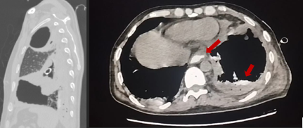
Fig 1:
He was taken up for left postero - lateral thoracotomy, about 1cm thick-walled empyema cavity was cut open, decortication was done. After mobilizing the left lung and mediastinum, a small rent of 5mm size in the lower thoracic Esophagus at the level of D10 on the postero medial wall was identified and repaired with 4-0 prolene sutures. The defect site was reinforced with an Omental patch after mobilizing and traversing esophageal hiatus. Intercostal drainage tube was kept in the left hemithorax. Feeding jejunostomy was done through a mini laparotomy by Gastrosurgery Department.
Post-operatively the patient was managed with brad spectrum intravenous antibiotic till the culture report arrived. In the initial days intermittent fever 103⁰F and raised inflammatory markers On POD 7, gastrograffin contrast Oesophagogram was repeated and leak was ruled out (fig 2). Oral sips and liquid were started on POD-8 which he gradually tolerated within 2 days following which the nasogastric tube and apical ICD were out.
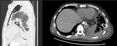
fig 2:
He was discharged on POD 11 with feeding jejunostomy and oral fluids in generally good condition with regular biweekly follow up with satisfactory chest radiograph. One month later, his oral intake was normal; feeding jejunostomy was removed.
Discussion
Boerhaave’s syndrome is a rare critical condition of spontaneous emetogenic esophageal perforation which is associated with high mortality and morbidity. Of all the esophageal perforation, this accounts for 15% only.
The pathogenic mechanism of Boerhaaves syndrome is due to a sudden rise in intraluminal oesophageal pressure against a closed glottis during vomiting. This is due to failure of cricopharyngeous muscle to relax. Neuromuscular coordination defect may be attributed to age and alcohol . Due to its anatomical structure, perforation of the oesophagus has a higher chance of occurring at four predisposing positions: 1) the Killian-Triangle; a muscle free zone in the proximal third of the oesophagus, 2) the crossing at the aortic arch, 3) the connection to the left main bronchus and 4) the esophagogastric junction. The most common site of perforation is on the left lateral wall just above the GEJ in two third of the patients as is seen in our case.
The classical triad of the Boerhaave’s syndrome include vomiting, chest pain and subcutaneous emphysema which is seen only in 14% cases. The time of patient presentation to hospital and correct diagnosis plays a crucial role in the outcome. The mortality is 10-25% if managed within 24 hours but it may reach as high as 60% when delayed and 100% if not intervened . The time is the utmost important as the mortality increases dramatically with delay in diagnosis. However, diagnosis and management are often delayed due to the rarity and presence of non-specific features. Misdiagnosis is the main concern in the management of this syndrome . Our patient was diagnosed the case on the day of admission, when he presented to us after 2 weeks.
On arrival to our hospital, a careful history taking which emphasized on an episode of forceful vomiting and subsequent onset of chest pain were the corner stone for considering the esophageal perforation. Adding to that, a persistent >1litre/day output in ICD was very unlikely of in the case of empyema. Provided late presentation and postprocedural status of the patient, Xray Chest and normal CECT thorax would provide very little information and hence we proceeded with Esophagogram with CECT thorax which confirmed the diagnosis.
We had two challenges. 1) a thorough debridement of the left hemithorax and mediastinum and 2) to find and close the esophageal defect.
When the patient presents within 24 hours and the wound is clean the management is primary repair of the rent, ICD and feeding jejunostomy. When presented after 24 hours and the wound bed is necrotic and debrided, then resection of diseased esophageal segment with cervical esophagostomy, stomach closure, ICD insertion and feeding jejunostomy. This is aimed at infection control, adequate nutrition and resuscitation. A second procedure after around 6 weeks is esophageal repair with sternal colon or stomach interposition.
Considering the difficulty of maintaining a cervical stoma and associated lifestyle adjustment, and a second procedure which can never be physiological and can cause lifelong morbidity, the young age with no comorbidity and reduce the financial burden, it was decided to proceed with a thorough debridement and primary closure of the perforation expecting a good recovery. Jacques et al in his studies in 27 cases, suggested long time interval does not pull back the surgeon from considering primary closure in Boerhaave's syndrome. But some authors are of the opinion that not all but some cases may benefit from the primary closure. A timely thoracic access , thorough decortication and meticulous multilayered closure at a well-equipped centres improve the outcome significantly. Even in delayed presentation, if the tissues are viable primary closure is suggested in some studies.
This is a rare case of a successful outcome of Boerhaave’s syndrome in spite of delayed presentation and high mortality rate. This is considered to be because of prompt and thorough evacuation of the septic focus, a feeding jejunostomy and nutrition. The corner stone was the perfect repair of the defect with reinforcement with omental patch, which is considered as The Abdominal Policeman. Omentum became the Surgeon’s golden patch due to several reasons; its highly vascular, promotes rapid healing of the repaired segment, enhances tissue perfusion and resists tissue ischemia, acts as a biological seal, a natural living buttress, has superior immune properties-controls infection, it’s a long and mobile pliable tissue which can be easily accessed as anyway an abdominal portal was created for feeding jejunostomy and there is expected low donor site morbidity.
After interventions of the esophageal rupture, the next crucial part of management for the patient’s prognosis is to control mediastinitis, intravenous antibiotic according to culture sensitivity, adequate drainage of mediastinum and pleural cavity, and high priority to nutrition was given.
Conclusion
Due to its rarity, early diagnosis of Boerhaave syndrome is the key for a better prognosis. Even in delayed presentation of Boerhaave syndrome, a skilful informed decision on primary repair helps to reduce the comorbidity, hospital cost, hospital stay in a well-equipped multi-speciality centre. A broader differential diagnosis from the history taking, which is the age-old practice, dying in the current era of high-end radio-guided treatment, should be kept in mind. It’s believed that the omental patch reinforcement of the oesophageal perforation repair helped in speedy wound healing.