- English
- عربي / Arabic
- മലയാളം / Malayalam

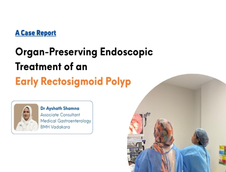
Advanced Endoscopic Management of an Early Rectosigmoid Neoplastic Polyp
12 August 2026Abstract
Colorectal polyps are recognized precursors of colorectal cancer, making early detection and complete endoscopic removal essential for preventing disease progression. We present the successful management of a young female patient with rectal bleeding who was diagnosed with a rectosigmoid adenomatous polyp. Initial colonoscopy identified the lesion, followed by staged therapeutic endoscopic resection. Histopathological examination demonstrated a tubulovillous adenoma with high-grade dysplasia and a minute focus of adenocarcinoma with superficial submucosal invasion (<1 mm). The patient underwent multidisciplinary evaluation with planned definitive surgical management. This case highlights the importance of high-quality endoscopy, timely histopathological diagnosis, multidisciplinary decision-making, and structured surveillance in achieving optimal patient outcomes.
Introduction
Colorectal cancer remains one of the leading causes of cancer-related morbidity and mortality worldwide. Most colorectal cancers develop gradually through the adenoma–carcinoma sequence, providing an opportunity for early diagnosis and intervention.
Advanced gastrointestinal endoscopy enables diagnosis and curative treatment of many premalignant lesions while preserving organs and reducing morbidity.
Patient Information and Clinical History
A 34-year-old female presented with intermittent bleeding per rectum for two days without abdominal pain. Digital rectal examination revealed blood staining without obvious anorectal pathology. She was referred to the Department of Gastroenterology for further evaluation.
Case Details
Complete colonoscopy after adequate bowel preparation demonstrated a rectal polyp approximately 5to 7 cm from the anal verge. Initial biopsy revealed tubular adenoma with low-grade dysplasia. Therapeutic sigmoidoscopy was subsequently performed, and a broad-based rectosigmoid polyp measuring approximately 2 cm was removed by piecemeal endoscopic polypectomy. No immediate bleeding or complications occurred. A scheduled re-look sigmoidoscopy confirmed healthy healing and no active bleeding. Final histopathology revealed tubulovillous adenoma with high-grade dysplasia and a small focus of adenocarcinoma with superficial submucosal invasion (<1 mm). The patient was referred for gastrointestinal surgical consultation for consideration of segmental colectomy.
Discussion
This case demonstrates the importance of investigating rectal bleeding irrespective of patient age. Complete endoscopic excision not only treated the visible lesion but also provided accurate pathological staging. Histopathology after complete removal revealed more advanced disease than the initial biopsy, emphasizing why complete excision is essential. Collaboration among gastroenterologists, endoscopy staff, pathologists and gastrointestinal surgeons ensured timely diagnosis and appropriate treatment planning.
Conclusion
Advanced endoscopic techniques played a pivotal role in early detection and management of this early colorectal neoplasm. The combination of expert colonoscopy, therapeutic polypectomy, histopathological evaluation and multidisciplinary care provided the patient with timely diagnosis and an excellent opportunity for curative treatment. This case reflects the commitment to evidence-based, minimally invasive gastrointestinal care.