A 47-year-old male with no history of any known comorbidities, sustained a cracker blast injury during a festival in a temple near Kozhikode. There was no initial history of breathlessness, hoarseness of voice, cough with soot, or carbonaceous sputum suggesting absence of overt inhalational injury.
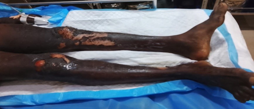
On arrival the patient was conscious and oriented. Immediate primary survey (ABCDE) was performed. On local examination approximately 50% TBSA mixed degree burns predominantly involving face, scalp, left upper limb and bilateral lower limbs noted. Singeing of fascial hairs and nasal hairs were present.
Initial Stabilization and Fluid Resuscitation
Initial management followed Advanced Burn Life Support (ABLS) principles. Airway, breathing, and circulation were assessed and stabilized. Elective intubation done. Fluid resuscitation was initiated using the Parkland formula (4 mL/kg/%TBSA), with half administered in the first 8 hours and the remainder over the next 16 hours. Urine output and hemodynamic parameters were closely monitored.
Wound Assessment, Surgical Debridement and Collagen Membrane Application
On PBD 1, the patient underwent thorough surgical debridement prior to biological wound coverage. Debridement was essential for removal of devitalized tissue, reduction of bacterial load, and accurate identification of deep dermal and full-thickness burn areas, facilitating early graft planning.
Following debridement, reassessment of burnt areas was done and found to have 50% TBSA second degree superficial to third degree burns involving face, scalp, left upper limb, bilateral lower limb and upper anterior trunk, with deep areas over bilateral lower limbs and left upper limb. Collagen membrane applied over the debrided areas and dried using warm air application. Collagen membranes act as temporary biological dressings, providing pain relief, reducing fluid loss, maintaining a moist wound environment, and aiding further demarcation of non-viable tissue.
Cadaveric Skin Grafting
Early excision and grafting constitute a cornerstone in the management of deep burn injuries.
Early excision and grafting within the first 3–7 days after burn injury is considered optimal for deep partial-thickness and full-thickness burns, once the patient is adequately resuscitated and hemodynamically stable. Prompt removal of devitalized and necrotic tissue reduces the bacterial load of the wound, thereby decreasing the risk of local infection and systemic sepsis, which remains a leading cause of mortality in burn patients. In addition, early grafting improves wound bed quality and enhances graft take, ultimately leading to better functional and aesthetic outcomes. In patients with extensive burns where immediate autografting is not feasible, early excision followed by temporary biological coverage, such as collagen membranes or cadaveric skin allografts, serves as an effective strategy to achieve these benefits while bridging the gap to definitive reconstruction.
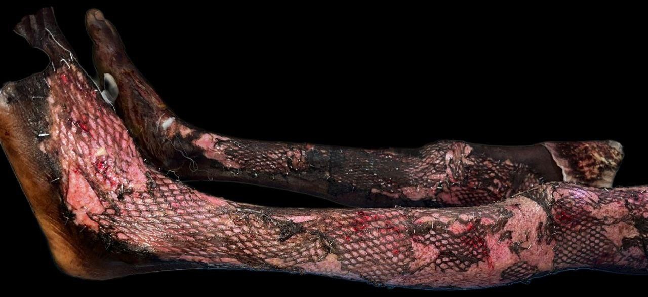
For our patient, due to the extensive burns and unavailability of donor areas, our next option for treatment of deep dermal and full-thickness burn areas was allografting with cadaveric skin. After an active search we could avail the required amount of cadaveric skin from an institute in Madurai, and grafting done on 05.01.2026 (PBD 6). After thorough debridement, tangential excision of deep areas were done till fresh bleed. Cadaveric skin applied over the excised areas and inset was given with skin staplers.
Postoperative care after cadaveric skin grafting is critical to ensure optimal graft adherence, prevent infection, and stabilize the patient until definitive wound coverage can be achieved. Since cadaveric skin acts as a temporary biological dressing, meticulous aftercare is essential to maximize its benefits.
Immediately after graft application, the grafted area is immobilized and covered with non-adherent sterile dressings to prevent shearing and displacement. Limb splintage is used where necessary to maintain graft stability. The first dressing change is usually delayed for 3–5 days unless there are signs of infection or excessive soakage.
The patient was closely monitored for local and systemic signs of infection. Broad-spectrum antibiotics are continued based on institutional protocol and modified according to culture sensitivity results. Regular assessment of graft adherence, color, exudate, and odor were performed to identify early graft failure or infection. Strict aseptic precautions were maintained during dressing changes.
The graft initially showed satisfactory take, followed by predictable immunological rejection. Despite rejection, significant dermal substance was preserved, improving readiness for definitive autografting and reducing scar formation.
Supportive Care
The hospital course was complicated by multiple infections:
Blood culture (5 January 2026): Grew Enterobacter cloacae
Pus culture (5 January 2026): Grew Klebsiella species
The patient received broad-spectrum and culture-directed antibiotics, including Amoxicillin-clavulanate, Piperacillin–tazobactam , Ciprofloxacin and Meropenem. The patient received broad-spectrum antibiotics, adequate analgesia, multiple blood transfusions, nutritional support, and meticulous monitoring for systemic complications. Clinical and laboratory parameters improved with therapy and was successfully extubated on postoperative day 1 following the second surgery.
Additional Supportive Care - Physiotherapy was initiated early to prevent joint stiffness and improve functional recovery. Persistent thrombocytosis was noted throughout the hospital stay, attributed to an inflammatory response secondary to extensive burns and infection, and was managed conservatively with close monitoring.
The patient showed gradual improvement in wound status and general condition. He was discharged on 20 January 2026 in a stable condition, with advice for continued wound care, physiotherapy, and outpatient follow-up.
Conclusion
Extensive burn injuries require a structured, staged approach to management in order to minimize early complications and optimize long-term outcomes. This case highlights the importance of early resuscitation, meticulous surgical debridement, and the strategic use of temporary biological dressings in managing major burns. The sequential application of collagen membrane followed by cadaveric skin grafting provided effective wound coverage, preserved dermal integrity, reduced fluid and protein losses, and facilitated wound bed preparation for definitive reconstruction. Although cadaveric skin serves only as a temporary cover and undergoes predictable immunological rejection, its role as a biological bridge in extensive burns is invaluable. This case reinforces the relevance of cadaveric skin grafting as an essential adjunct in modern burn care, particularly in patients with limited autograft availability, and underscores its role in improving wound management and overall patient stabilization.