- English
- عربي / Arabic
- മലയാളം / Malayalam

When the Parathyroid Hides in the Carotid Sheath
05 October 2026When the Parathyroid Hides in the Carotid Sheath
An unusual journey to cure primary hyperparathyroidism
The Clinical Clue
A 31-year-old woman presented with a four-year history of bone pain, recurrent upper abdominal pain and headaches. Investigations revealed bilateral renal calculi, nephrocalcinosis, corrected calcium of 11.3 mg/dL, markedly elevated parathyroid hormone (PTH), and a pepper-pot appearance on skull radiography.
A Small Gland, a Large Clinical Footprint
A 31-year-old woman reached our Endocrine Surgery department after several years of symptoms. Hypercalcaemia had been detected earlier, but surgery had been deferred after an ultrasound of the neck performed outside failed to localize a parathyroid lesion. Over time, the disease left its mark: bone pain persisted, and bilateral renal calculi with nephrocalcinosis developed.
Her biochemical profile was consistent with primary hyperparathyroidism. The challenge was no longer establishing the diagnosis-it was finding the abnormal gland.
Where Was the Culprit Gland?
The outside ultrasound had been negative. 99mTc-sestamibi scintigraphy, however, suggested a left inferior parathyroid lesion. A targeted ultrasound was then performed in our Department of Endocrine Surgery. It revealed a 13 × 11 × 8 mm hypoechoic lesion in the left lower neck, about 3 cm below the lower pole of the thyroid—lateral to the common carotid artery and medial to the internal jugular vein. The finding changed the surgical plan from uncertainty to a precisely directed exploration.
Following the imaging- even when the usual glands looked normal
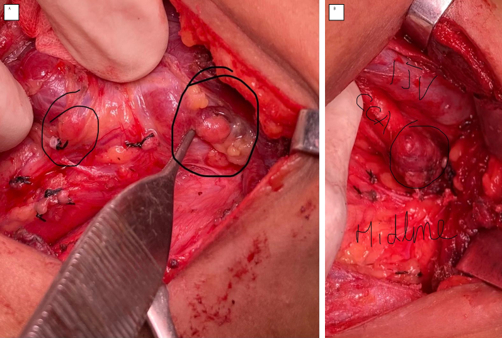
Operative photographs: (A) normal ipsilateral parathyroid glands identified in their expected positions; (B) the ectopic parathyroid adenoma within the carotid sheath, adjacent to the great vessels.
At exploration, both the left superior and left inferior parathyroid glands were found in their normal positions and appeared normal.
• The carotid sheath was carefully opened.
• The vagus nerve was identified and safeguarded.
• A fleshy 11 × 13 × 10 mm lesion was found over the carotid artery, medial to the internal jugular vein.
• The lesion was meticulously excised and sent for histopathology.
• Ten minutes after excision, intraoperative PTH fell to 26 pg/mL—confirming successful removal.
Why can a parathyroid be found there
Parathyroid glands travel during embryological development, and a variation in that journey can leave a gland far from its expected position. Ectopic glands may lie in the thymus, mediastinum, retro- or para-oesophageal region, within the thyroid, or - much more rarely - in the carotid sheath.
Why this case stands out
Carotid-sheath parathyroid adenomas are exceptionally uncommon. In the literature reviewed for the original report, only 18 adenomas had been described in or around the carotid sheath, and only nine were actually within the sheath. Some reported patients required repeated explorations before the culprit gland was found; one historical case was cured only after five operations. In contrast, this patient was localized accurately and cured at the first operation.
The patient recovered well and was discharged on postoperative day 2 without clinical hypocalcaemia.
The best localization for a difficult parathyroid is to locate the right parathyroid surgeon - one who can combine biochemistry, imaging, embryology and operative anatomy to find the gland safely.
A PARATHYROID SURGEON’S PERSPECTIVE A negative ultrasound does not end the search. The biochemical diagnosis comes first; imaging is used to localize the abnormal gland. Ultrasound is operator-dependent, and a targeted study by an experienced team can reveal a lesion missed elsewhere. When both expected ipsilateral glands look normal, the preoperative localization should still be respected. In this case, opening the carotid sheath, identifying and protecting the vagus nerve, and confirming cure with intraoperative PTH converted a rare anatomical surprise into a successful first operation.