- English
- عربي / Arabic
- മലയാളം / Malayalam

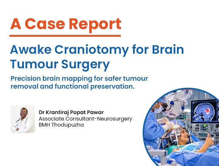
Awake Craniotomy: Preserving Speech and Movement
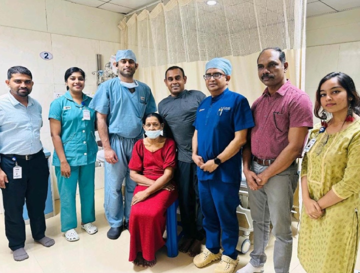
27 October 2026In a significant milestone for advanced neurosurgery, our medical team has successfully performed a complex awake craniotomy to resect a high frontal Intracranial Space-Occupying Lesion (ICSOL) located within the brain's critical eloquent cortex. The patient, who faced a high risk of permanent speech and motor deficits due to the tumor's delicate location, has shown exceptional postoperative recovery with all neurological functions perfectly preserved.
This surgical achievement underscores the power of modern mapping technologies, specialized neuro-anesthesia, and elite surgical precision coming together to turn a highly precarious diagnosis into a triumphant story of clinical success.
An Intracranial Space-Occupying Lesion (ICSOL) refers to any abnormal mass inside the cranium, such as a primary brain tumor, metastasis, or cyst, that increases intracranial pressure and displaces normal brain tissue. When a tumor presents in the left high frontal lobe, the stakes are exceptionally high. In the vast majority of individuals, the left hemisphere is the dominant side of the brain, housing critical networks responsible for language production, expressive speech, motor control, and higher-order executive functioning.
The specific region surrounding this patient's tumor is classified as an eloquent area—a zone of the brain where any surgical damage results in immediate, irreversible neurological deficits, such as a complete loss of speech (aphasia) or right-sided paralysis (hemiparesis).
Traditional neurosurgery under general anesthesia relies on structural landmarks and pre-operative imaging. However, every individual's brain wiring is unique. When a tumor infiltrates or compresses eloquent tissue, conventional surgery poses an unacceptable risk of clipping vital pathways. To maximize tumor removal while entirely safeguarding the patient's personality, language, and movement, the surgical team chose the gold standard for eloquent zone surgery: an awake craniotomy.
An awake craniotomy is an ultra-specialized procedure where the patient is deliberately awakened midway through surgery while the neurosurgeon operates on the brain. Because the brain tissue itself does not possess pain receptors, the patient experiences no physical discomfort from the surgical resection once the scalp and skull are properly numbed.
The primary objective of keeping the patient awake is to perform intraoperative neurocognitive mapping. This technique allows the surgical team to interact directly with the patient in real-time. By testing speech, comprehension, memory, and motor functions while precisely stimulating the brain tissue surrounding the tumor, surgeons can map out an individualized "no-fly zone." If a specific area is stimulated and the patient experiences a transient speech arrest or muscle weakness, the surgeon instantly knows that tissue is functionally vital and must be preserved.
This real-time feedback loop removes the guesswork from complex tumor resections. It shifts the surgical paradigm from an anatomical approach to a functional one, pushing the boundaries of how much of a tumor can be safely removed.
The success of this breakthrough case relied heavily on a seamless, multi-disciplinary approach involving neurosurgeons, neuro-anesthesiologists, clinical neurophysiologists, and specialized nursing teams. The procedure was executed using the sophisticated Asleep-Awake-Asleep (AAA) protocol.
1. The Pre-Operative Preparation and First "Asleep" Phase
The journey began weeks before the operating room, where the patient underwent rigorous clinical psychological conditioning. The patient was coached on what to expect, the sounds of the operating room, and the specific language and motor tasks they would perform mid-surgery.
On the day of the operation, advanced neuro-anesthesiologists administered tailored, short-acting intravenous sedatives. This kept the patient completely asleep, comfortable, and pain-free during the initial phases of the surgery, which included securing the head frame, performing the scalp incision, and creating the bone flap (the craniotomy) to expose the left high frontal lobe.
2. The Critical "Awake" Phase: Real-Time Cortical Mapping
Once the brain surface was safely exposed and the surgical field stabilized, the anesthesiologist carefully titrated down the sedation, bringing the patient to a fully conscious, cooperative, and interactive state within minutes.
With the patient awake and comfortable under regional scalp blocks, the intraoperative mapping began. The neurophysiologist stood at the patient’s side, utilizing flashcards, verbal exercises, and finger-movement protocols. Simultaneously, the neurosurgeon used a specialized, low-current bipolar electrical stimulator to gently touch areas of the brain adjacent to the left high frontal ICSOL.
As the patient counted aloud or named objects on a screen, the surgeon observed the effects of the micro-stimulations. When stimulation near the lower border of the tumor caused a brief pause in the patient's speech, that exact coordinate was marked as functionally eloquent. This mapped a safe, customized surgical corridor directly to the tumor, completely avoiding vital speech pathways (such as Broca's area) and primary motor tracts.
Using high-magnification neurosurgical microscopes and ultrasonic aspirators, the surgeon meticulously peeled the tumor away from the healthy, mapped brain tissue. Throughout the entire resection, the patient continued to converse and perform motor tasks, providing a continuous, real-time safety report.
3. The Final "Asleep" Phase: Closure
Once the maximal safe resection of the high frontal mass was achieved, the patient was gently placed back under general anesthesia. The surgical team then completed the meticulous closure of the dura mater, replaced the bone flap securely with titanium microplates, and sutured the scalp incision.
We are thrilled to report that the postoperative outcome has been outstanding. Immediately following the surgery, the patient was transferred to the Neuro-Intensive Care Unit for close observation. Neurological assessments conducted just hours after the procedure confirmed that the patient’s speech fluency, word recall, comprehension, and motor strength on the right side of the body remained fully intact.
Because an awake craniotomy avoids the prolonged use of heavy, deep general anesthetics, patients experience significantly less postoperative nausea, cognitive clouding, and systemic fatigue. This patient was sitting up, eating, and interacting with family members by the very next day.
Follow-up neuroimaging has confirmed an excellent extent of resection, showing maximum clearance of the mass while completely sparing the surrounding healthy brain framework. The patient has since been discharged home in excellent spirits, continuing their path toward a full return to daily life with their quality of life entirely preserved.
This successful outcome highlights the immense clinical value of awake craniotomies for tumors involving eloquent regions. Historically, tumors invading the dominant left frontal lobe were often deemed inoperable, or were resected conservatively, leaving significant residual tumor tissue behind out of fear of causing permanent disability.
By leveraging intraoperative functional mapping, our neurosurgical department effectively minimizes these risks. This breakthrough demonstrates that with the right combination of surgical expertise, technological infrastructure, and compassionate patient preparation, even the most daunting neurological challenges can be navigated safely.
Our center remains deeply committed to pioneering these advanced neurosurgical interventions, ensuring that our patients receive world-class, precise, and deeply individualized care when they need it most.